
Difference between Ultrasound and Sonogram
Key Difference: In common parlance, ultrasounds and sonograms are essentially synonymous. They are a medical test which uses high frequency sound waves to produce a picture of the organs in the body. In a strict technical sense, ultrasound refers to the actual frequency above that which humans can hear. Sonography, on the other hand, technically refers to the practice of using the ultrasound frequency to create images inside the body.
 In common parlance, ultrasounds and sonograms are essentially synonymous. They are a medical test which uses high frequency sound waves to produce a picture of the organs in the body. The process is called an ultrasound scan, whereas the images are often referred to as sonograms. An ultrasound scan may also be referred to as a sonogram, diagnostic sonography, and ultrasonography.
In a strict technical sense, ultrasound refers to the actual frequency above that which humans can hear. The limit is generally considered to be 20 kilohertz (20,000 hertz) in healthy, young adults. Ultrasound devices operate with frequencies from 20 kHz up to several gigahertz. Sonography, on the other hand, technically refers to the practice of using the ultrasound frequency to create images inside the body. Sonography is also commonly called diagnostic sonography or ultrasonography.
Ultrasound is used in many different fields. It can be used for imaging, detection, measurement, and cleaning. Ultrasonics, i.e. the application of ultrasound, is also useful for changing the chemical properties of substances at higher power levels.
Medically, ultrasound is used to visualizing body structures using an ultrasound-based diagnostic imaging technique. It can be used to visualize body structures such as tendons, muscles, joints, vessels and internal organs. The images gained can be used to look for and diagnose possible pathology or lesions.
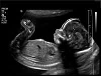
During pregnancy, ultrasounds are used to produce an image of the fetus. According to the American Pregnancy Association, there are seven types of ultrasounds: transvaginal, standard (2 dimensional or 2D), advanced (targeting a specific issue of concern), fetal echocardiography (assessing the heart), Doppler, 3D and 4D (also known as “dynamic” 3D because it focuses on the face and fetal movements). Ultrasounds are usually done in order to keep track of the pregnancy and to ensure that the baby is safe and healthy. 3D Ultrasound and 4D Ultrasounds have gained a lot of popularity with expecting parents.
Image Courtesy: upmc.com
 |
 |
 |
 |
 |
 |
 |
 |
Add new comment